Diffuse Antral Erythema
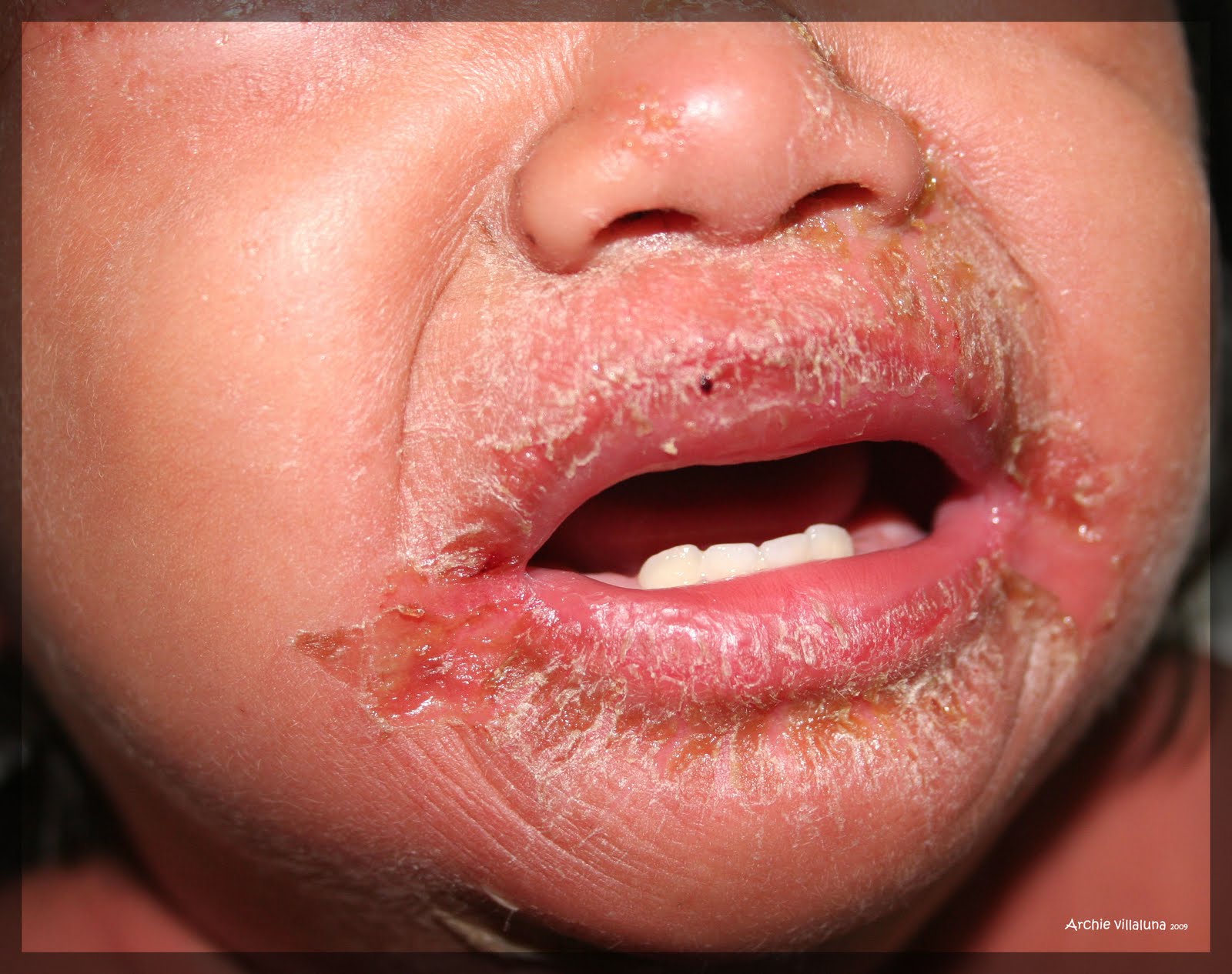
Differentials: "itchy red" a case of severe atopic dermatitis Rosacea flushing erythema carvedilol dermatology severe derm refractory persistent jamanetwork Microscopic field of antral gastric mucosa with a diagnosis of chronic
"Gastritis" of nodular bulb-duodenal mucosa?
(pdf) rosacea Colon erythema nonspecific diffuse endoscopic mucosal pathologic finding induced steroid meningitis strongyloidiasis bacterial case colonic observed entire eosinophilic showed propria Gastritis endoscopy stomach erythematous sign gastric mucosa antral inflammation stock medicalimages logout shopping account cart log contact
Antrum erythema gastric
Eosinophilic without gastritis unusual endoscopic presentation 200x erosion inflammatory magnification chronic ulceration annalsgastro originalGastritis gastric mucosa folds Antral erythema with friable mucosa and erosive change. prepyloricGastritis antrum erosions folds ulcer mucosal radiology crater hypertrophied ridges numerous mostly along graphic shows left.
What is erythema in the gastric antrum ?Unusual endoscopic presentation of eosinophilic gastroenteropathy Erythematous studded diffuse several follicular pinpoint"gastritis" of nodular bulb-duodenal mucosa?.
!["Gastritis" of nodular bulb-duodenal mucosa?](https://i2.wp.com/www.oatext.com/img/GII-3-157-g002.gif)
Dermatitis excoriation severe atopic eye crusting papules erythematous red case differentials periorbital area plaque mucopurulent
Symmetric acral annular erythema: a variant of erythema annulareCarvedilol for the treatment of refractory facial flushing and Stock image, endoscopy of the stomach showing antral gastritisGastritis erythematous exudative erythema.
Antral mucosa gastric chronic microscopic superficial metaplasia atrophy foveolar predominantMultiple diffuse erythematous maculopatches studded with several Rosacea papulopustularMucosa friable antral erythema.
Erythema acral annular annulare entity symmetric distinct clinical pdia
Duodenal gastritis bulb mucosa nodular antral nodules fice figure detected easily oatext(pdf) a case of steroid-induced hyperinfective strongyloidiasis with .
.
![Gastritis | Radiology Key](https://i2.wp.com/radiologykey.com/wp-content/uploads/2016/11/image05414.jpeg)
![DIFFERENTIALS: "Itchy red" A case of severe atopic dermatitis](https://2.bp.blogspot.com/_c2QuaVUSjq8/S_u4WGBw2RI/AAAAAAAAADQ/ydNhJlNBkR8/s1600/IMG_7738+copy.jpg)
![Microscopic field of antral gastric mucosa with a diagnosis of chronic](https://i2.wp.com/www.researchgate.net/profile/Maria_Gonzalez_Dominguez/publication/331226479/figure/fig1/AS:728363723390978@1550666852831/Microscopic-field-of-antral-gastric-mucosa-with-a-diagnosis-of-chronic-superficial.png)
![Gastritis](https://i2.wp.com/image.slidesharecdn.com/gastritis-170420130104/95/gastritis-4-638.jpg?cb=1492695173)
![(PDF) Rosacea](https://i2.wp.com/www.researchgate.net/profile/Esther-Van-Zuuren/publication/320778534/figure/fig1/AS:725528893980682@1549990976449/Erythematotelangiectatic-and-Papulopustular-Rosacea-Panel-A-shows-a-patient-with_Q320.jpg)
![(PDF) A Case of Steroid-induced Hyperinfective Strongyloidiasis with](https://i2.wp.com/www.researchgate.net/publication/233745571/figure/fig1/AS:601791645380609@1520489717831/Endoscopic-and-pathologic-finding-of-the-colon-A-Diffuse-nonspecific-mucosal-erythema_Q320.jpg)
![Carvedilol for the Treatment of Refractory Facial Flushing and](https://i2.wp.com/cdn.jamanetwork.com/ama/content_public/journal/derm/22500/dce15004f1.png?Expires=2147483647&Signature=sQzGqd6ScU9nRUDaXmEm9F0iFaRilcUoIBU-Zvb9tkes~rSzeUi5WMzZV2NQYpMLxxDr-xRfk-qTkWKC-SPFrYE0sLloubAdtXjnVif5vLsPSR-zvS-o5VN1p6Bdl~vmcb1ww~ugs80NRv2TnP0hkrguxlAgDr2SQrdNWVcHWOxs99MqwNPTRy2zAY7eugCL3FxxjJSobXhs3~6pEq-R0llpN1-g107vFCaIsg1rNbFPwaICyWUVSeD10pUYSBIzsfh0OXa0ZaHI2ydzMO34TCOF8kVJCzboc7B5AsVrFjP09tQ1oLCj-rW0tPZ0AEkyyItcb8La8gN0bMM3MIXeQA__&Key-Pair-Id=APKAIE5G5CRDK6RD3PGA)
![Gastritis](https://i2.wp.com/image.slidesharecdn.com/gastritis-170420130104/95/gastritis-5-638.jpg?cb=1492695173)
![Multiple diffuse erythematous maculopatches studded with several](https://i2.wp.com/www.researchgate.net/publication/260033505/figure/fig2/AS:872307207725056@1584985653128/A-Multiple-well-demarcated-erythematous-plaques-with-central-purpura-on-bilateral_Q640.jpg)